• Characterization of di-4-ANEPPS with nano-black lipid membranes
    M. Tsemperouli and K. Sugihara
    Nanoscale, in press (2017)
    DOI:10.1039/C7NR05863B | Abstract | Article PDF | Supporting Info
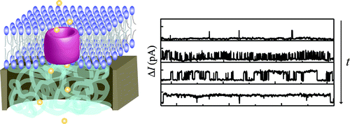
We report a platform based on lateral nano-BLMs, where electrical measurements and fluorescence microscopy setup are combined, for the calibration of di-4-ANEPPS, a common voltage sensitive dye (VSD). The advantage of the setup is 1) its flexibility in the choice of lipids and the applied voltages, 2) its high stability that enables high voltage (500 mV) application and long time measurements, and 3) its fluorescence microscopy readout, which can be directly correlated with other fluorescence microscopy experiments using VSDs (e.g. membrane potential measurements in living cells). Using the setup, we observed that the calibration curve of di-4-ANEPPS highly depends on the net electric charge of the lipids. The developed setup can be used to calibrate VSDs in different lipid environment for understanding their fundamental voltage-sensing mechanism in future.
  • Artificial tubular connections between cells based on synthetic lipid nanotubesOpen access paper
    A. Kozintsev and K. Sugihara
    RSC Advances, 7 (33) (2017), p20700-20708
    DOI:10.1039/C7RA02187A | unige:94060 | Abstract | Article HTML | Article PDF
 
Tunneling nanotubes (TNTs) have become a major topic of interest as a form of intercellular communication due to their recent discovery. However, research on this subject has often suffered from a lack of controllability in the generation of the nanotubular connections. In this work, we demonstrate a simplified approach to selectively create a direct nanotubular connection between eukaryotic cells by manually manipulating self-assembling lipid nanotubes (LNTs) from inverted hexagonal-phase lipid blocks. The technique requires minimal instrumentation for creating the LNT connection between cells compared to conventional approaches. Based on the diffusion of fluorescent lipids from LNTs into cell membranes (D = 0.032 ± 0.003 μm2 s−1), the probability of observing membrane fusion between LNTs and cell membranes was estimated as 30%. Among these cell–LNT junctions the resulting structure is open-ended roughly 75% of the time, as evidenced from observations of the diffusion of a water-soluble dye between two cells connected with this nanotubular structure.
  
  • Mechanosensitivity of polydiacetylene with a phosphocholine headgroup
    R.D. Ortuso, U. Cataldi and K. Sugihara
    Soft Matter, 13 (8) (2017), p1728-1736
    DOI:10.1039/C6SM02579J | unige:92341
We demonstrated colorimetric and fluorescence detection of peptide, melittin, with polydiacetylene (PDA) made of 1,2-bis(10,12-tricosadiynoyl)-sn-glycero-3-phosphocholine (DC(8,9)PC). The PDA used in this work has a phosphocholine headgroup, which mimics peptide-cell membrane interactions better than the conventional PDA assays with carboxyl headgroup. The dose curve (colorimetric response vs melittin concentration) showed a half maximum response at the melittin concentration of 0.1 mg/ml, which is similar to the traditional PDA assays. It suggests that the replacement of the headgroup was achieved without sacrificing the sensitivity. From the dose curve, Hill coefficient was extracted as αHill = 2.1. The value is in agreement with previous melittin studies with standard phospholipids, which reflects the benefit of having a biologically relevant headgroup. In addition, we found an unexpectedly slow spectral change when DC(8,9)PC-PDA was incubated with melittin. The origin of the time-dependent signal was studied by combining UV/VIS spectroscopy, fluorescence spectroscopy and dynamic light scattering.
  • Self-assembled Lipid Structures as Model Systems for Studying Electrical and Mechanical Properties of Cell Membranes
    K. Sugihara
    Chimia, 70 (11) (2016), p805-809
    DOI:10.2533/chimia.2016.805 | unige:90820 | Abstract
This mini review summarizes our recent works in the development of electrical and mechanical characterization tools for cell membranes. These research topics require the application of physical and chemical characterization tools to biological systems, thus are very interdisciplinary.
  • Gold Nanowire Fabrication with Surface-Attached Lipid Nanotube Templates
    K. Jajcevic, M. Chami and K. Sugihara
    Small, 12 (35) (2016), p4830-4836
    DOI:10.1002/smll.201600431 | unige:87903 | Abstract | Article HTML | Article PDF

A high-throughput approach to fabricate gold nanowires on surfaces with a lipid nanotube template is demonstrated. Streptavidin-coated gold nanoparticles are attached to the biotin-tagged lipid nanotubes. After the chemical fixation, the samples are dried and treated with oxygen plasma to remove the organic template and connect the particles. The created nanowires are characterized by cryo-transmission electron microscopy, atomic force microscopy, and electrical measurements.
 
Polydiacetylene (PDA) is a conductive polymer that has a mechanochromism. When the polymer is exposed to mechanical stresses, change in temperature (thermochromism), pH (ionochromism) etc., the structural perturbation can be seen by the change in its color. Although it presents interesting electrical and optical properties, the relationship between these signals has rarely been investigated. We studied the correlation between the electrical conductivity and the absorption spectra of PDA. Upon UV irradiation, PDA absorption spectra presented a blue shift, which coincided with the decrease in the electrical conductivity.
LNTs are unique 3D structures made only of safe and abundant biomaterials by self-assembly. The current bottleneck for developing applications using LNTs is the lack of an easy technique to pattern them on substrates. We report a method to free-draw single lipid nanotube (LNT) patterns in any shape on surfaces with 1,2-dioleoyl-sn-glycero-3-phosphoethanolamine (DOPE) that takes an inverted hexagonal (HII) phase. We used pre-self-assembled LNTs or HII lipid blocks as a lipid reservoir from which new LNTs were pulled by applying a point load with a micromanipulator. The extreme simplicity of our technique originates from the fundamental nature of DOPE lipids that prefer a HII phase, while all the conventional approaches use PC lipids that form a lamellar phase. By adjusting the surface properties with polyelectrolyte multilayers, the created single LNT objects are able to remain adhered to the surface for over a week. Importantly, it could be shown that two vesicles loaded with caged fluorescent molecules were able to fuse well with a LNT, enabling diffusive transport of uncaged fluorescent molecules from one vesicle to the other.
  • Artificial Bacterial Flagella for Remote-Controlled Targeted Single-Cell Drug Delivery
    R. Mhanna, F. Qiu, L. Zhang, Y. Ding, K. Sugihara, M. Zenobi-Wong and B.J. Nelson
    Small, 10 (10) (2014), p1953-1957
    DOI:10.1002/smll.201303538 | Abstract | Article HTML | Article PDF | Video S1
An approach for batch preparation of liposome-functionalized microdevices is demonstrated for remotely controlled single-cell drug delivery. The liposome functionalized artificial bacterial flagella exhibit corkscrew swimming in 3D with micrometer positioning precision by applying an external rotating magnetic field. The devices are also capable of delivering water-soluble drugs to single cells in vitro.
  • Artificial bacterial flagella functionalized with temperature-sensitive liposomes for biomedical applications
    F. Qiu, R. Mhanna, L. Zhang, Y. Ding, K. Sugihara, M. Zenobi-Wong and B.J. Nelson
    in "Solid-State Sensors, Actuators and Microsystems (TRANSDUCERS & EUROSENSORS XXVII)" 2013 Transducers & Eurosensors XXVII: The 17th International Conference on, 27 (2013), p2130-2133
    DOI:10.1109/Transducers.2013.6627222 | Abstract | Article HTML | Article PDF
Inspired by flagellar propulsion of bacterial such as E. coli, artificial bacterial flagella (ABFs) are magnetic swimming microrobots with helical shapes. ABFs can perform precise three-dimensional (3D) navigation in liquids under low-strength rotating magnetic fields making them attractive tools for drug delivery applications. Further functionalization of these swimming microrobots is necessary to optimize their performance of biomedical tasks. We report here for the first time the successful functionalization of titanium-coated ABFs with temperature-sensitive dipalmitoylphosphatidylcholine (DPPC) liposomes. Adsorption of intact liposomes on titanium was assessed using quartz crystal microbalance with dissipation monitoring (QCM-D). The adsorption of fluorescently labeled liposomes on the surface of ABFs was confirmed with confocal laser scanning microscopy (CLSM) images. Functionalized ABFs (f-ABFs) can be loaded with both hydrophilic and hydrophobic drugs, and controlled drug release triggered by temperature. ABFs have a great potential to be used in targeted and controlled drug delivery and for in vivo sensing.
  
  • Switching Transport through Nanopores with pH-Responsive Polymer Brushes for Controlled Ion Permeability
    G.W. De Groot, M.G. Santonicola, K. Sugihara, T. Zambelli, E. Reimhult, J. Vörös and G.J. Vancso
    ACS Applied Materials & Interfaces, 5 (4) (2013), p1400-1407
    DOI:10.1021/am302820y | Abstract | Article HTML | Article PDF
Several nanoporous platforms were functionalized with pH-responsive poly(methacrylic acid) (PMAA) brushes using surface-initiated atom transfer radical polymerization (SI-ATRP). The growth of the PMAA brush and its pH-responsive behavior from the nanoporous platforms were confirmed by scanning electron microscopy (SEM), Fourier transform infrared (FTIR) spectroscopy, and atomic force microscopy (AFM). The swelling behavior of the pH-responsive PMAA brushes grafted only from the nanopore walls was investigated by AFM in aqueous liquid environment with pH values of 4 and 8. AFM images displayed open nanopores at pH 4 and closed ones at pH 8, which rationalizes their use as gating platforms. Ion conductivity across the nanopores was investigated with current–voltage measurements at various pH values. Enhanced higher resistance across the nanopores was observed in a neutral polymer brush state (lower pH values) and lower resistance when the brush was charged (higher pH values). By adding a fluorescent dye in an environment of pH 4 or pH 8 at one side of the PMAA-brush functionalized nanopore array chips, diffusion across the nanopores was followed. These experiments displayed faster diffusion rates of the fluorescent molecules at pH 4 (PMAA neutral state, open pores) and slower diffusion at pH 8 (PMAA charged state, closed pores) showing the potential of this technology toward nanoscale valve applications.
  • Label-free detection of cell-contractile activity with lipid nanotubes
    K. Sugihara, M. Delai, R. Mahnna, J. Kusch, D. Poulikakos, J. Vörös, T. Zambelli and A. Ferrari
    Integrative Biology, 5 (2) (2013), p423-430
    DOI:10.1039/c2ib20202f | Abstract | Article HTML | Article PDF
 
Surface-bound self-assembled lipid nanotubes (LNTs) made of 1,2-dioleoyl-sn-glycero-3-phosphoethanolamine (DOPE) were used to visualize the contractile activity of spreading cells. The interaction of cells with LNTs resulted in the nucleation of new nanotubes, directed toward the cell center, from existing ones. This process depended on cell generated forces and required acto-myosin mediated contractility. The dynamics of de novo generation of LNTs upon cell spreading was captured using optical microscopy on fluorescently labeled nanotubes and revealed characteristic fingerprints for different cell types such as fibroblasts, endothelial and melanoma cells. Additionally, the method was applied to detect the effect of a specific inhibitor on the generation of cellular forces. The mechanism of the LNT–cell interaction and the potential applications are discussed.
  
  • Directed Self-Assembly of Lipid Nanotubes from Inverted Hexagonal Structures
    K. Sugihara, M. Chami, I. Derényi, J. Vörös and T. Zambelli
    ACS Nano, 6 (8) (2012), p6626-6632
    DOI:10.1021/nn300557s | Abstract | Article HTML | Article PDF
Conventional lipid-tube formation is based on either a tube phase of certain lipids or the shape transformation of lamellar structures by applying a point load. In the present study, lipid blocks in inverted hexagonal phase made of 1,2-dioleoyl-sn-glycero-3-phosphoethanolamine (DOPE) were shown to protrude lipid nanotubes upon a fluid-dynamic flow on polyelectrolyte-functionalized surfaces in physiological buffer solution. The outer diameter of the tubes is 19.1 ± 4.5 nm and their lengths are up to several hundred micrometers. The method described enables the alignment and patterning of lipid nanotubes into various (including curvy) shapes with a microfluidic system.
 
Inverted hexagonal blocks of 1,2-dioleoyl-sn-glycero-3-phosphoethanolamine (DOPE) lipid adsorbed on a polyethyleneimine (PEI)-coated surface in deionized water transformed its shape upon the application of an electric field, forming lipid objects in a variety of shapes (e.g. lines with a width of 10–50 μm). The phenomenon was driven by the electrophoresis, because the zwitterionic lipid, DOPE turned out to be highly negatively charged in deionized water. The interaction between DOPE and the PEI surface stabilized the system, assuring a lifetime over several weeks for the formed structures after the electric field was switched off. The free-drawing of microscopic objects (lines, crosses, and jelly fish) was also achieved by controlling the direction of the lipid movement with the field direction.
  
  • A universal method for planar lipid bilayer formation by freeze and thaw
    K. Sugihara, B. Jang, M. Schneider, J. Vörös and T. Zambelli
    Soft Matter, 8 (20) (2012), p5525-5531
    DOI:10.1039/c2sm25148e | Abstract | Article HTML | Article PDF
A procedure based on freezing and thawing was developed to induce the rupture of adsorbed lipid vesicles on solid surfaces into supported lipid bilayers (SLBs). The SLB assembly exploits the phase transition of both lipids and water during freezing. It enables SLB formation independent of the type of substrates and lipids as long as the vesicles spontaneously adsorb onto the surface. The created SLB is a single bilayer, and has a diffusion coefficient of (0.6–4) × 10−8 cm2 s−1 on TiO2, which is in the same range as the SLBs formed by conventional techniques. The presented approach has the advantages of both the Langmuir–Blodgett method (the versatility in the selection of lipids and substrates) and vesicle fusion (self-assembly) at the same time.
The enormous progress of nanotechnology during the last decade has made it possible to fabricate a great variety of nanostructures. On the nanoscale, metals exhibit special electrical and optical properties, which can be utilized for novel applications. In particular, plasmonic sensors including both the established technique of surface plasmon resonance and more recent nanoplasmonic sensors, have recently attracted much attention. However, some of the simplest and most successful sensors, such as the glucose biosensor, are based on electrical readout. In this review we describe the implementation of electrochemistry with plasmonic nanostructures for combined electrical and optical signal transduction. We highlight results from different types of metallic nanostructures such as nanoparticles, nanowires, nanoholes or simply films of nanoscale thickness. We briefly give an overview of their optical properties and discuss implementation of electrochemical methods. In particular, we review studies on how electrochemical potentials influence the plasmon resonances in different nanostructures, as this type of fundamental understanding is necessary for successful combination of the methods. Although several combined platforms exist, many are not yet in use as sensors partly because of the complicated effects from electrochemical potentials on plasmon resonances. Yet, there are clearly promising aspects of these sensor combinations and we conclude this review by discussing the advantages of synchronized electrical and optical readout, illustrating the versatility of these technologies.
  • Simultaneous OWLS and EIS monitoring of supported lipid bilayers with the pore forming peptide melittin
    K. Sugihara, M. Delai, I. Szendro, O. Guillaume-Gentil, J. Vörös and T. Zambelli
    Sensors and Actuators B: Chemical, 161 (1) (2012), p600-606
    DOI:10.1016/j.snb.2011.11.007 | Abstract | Article HTML | Article PDF
 
A simultaneous optical waveguide lightmode spectroscopy (OWLS) and electrochemical impedance spectroscopy (EIS) measurement was carried out for the investigation of a supported lipid bilayer and its interactions with a pore-forming peptide, melittin. It was achieved only after the optimization of the ITO coating on the waveguide to increase the electrical sensitivity and the functionalization of the waveguide with a polyelectrolyte to form a lipid bilayer over the ITO surface. The combined system enabled monitoring of melittin pore activities in a wider range of melittin concentrations than either technique alone (1 μg/ml < Cmelittin < 200 μg/ml). Furthermore, it provided unique information that could not be obtained by the individual methods, such as a better identification of the melittin-pore formation and an insight about the correlation between the total pore area vs. adsorbed amount of melittin.
  • Techniques for recording reconstituted ion channels
    S. Demarche, K. Sugihara, T. Zambelli, L. Tiefenauer and J. Vörös
    Analyst, 136 (6) (2011), p1077-1089
    DOI:10.1039/c0an00828a | Abstract | Article HTML | Article PDF
This review describes and discusses techniques useful for monitoring the activity of protein ion channelsin vitro. In the first section the biological importance and the classification of ion channels are outlined in order to justify the strong motivation for dealing with this important class of membrane proteins. The expression, reconstitution and integration of recombinant proteins into lipid bilayers are crucial steps to obtain consistent data when working with ion channels. In the second section recording techniques used in research are presented. Since this review focuses on analytical systems bearing reconstituted ion channels the industrial most important patch-clamp techniques of cells are only briefly mentioned. In section three, artificial systems developed in the last decades are described while the emerging technologies using nanostructured supports or microfluidic systems are presented in section four. Finally, the remaining challenges of membrane protein analysis and its potential applications are briefly outlined.
  
The resistivity ÏPEM of polyelectrolyte multilayers (PEMs), PEI(PSS/PAH)24, PEI(PGA/PAH)12, PEI(HA/PLL)12 and PEI(PSS/PLL)12, in a free-hanging configuration was estimated combining electrochemical impedance spectroscopy (EIS) and atomic force microscopic (AFM) images. Surprisingly, the obtained value of several kΩcm is at least 6 orders of magnitude lower than that reported previously, where the resistivity was determined in the conventional PEM-on-electrode system. The significant discrepancy indicates the unexpectedly low electrical PEM resistance in the absence of redox-active ions and the sensitivity limitation in the conventional system.
  • Liposome and Lipid Bilayer Arrays Towards Biosensing Applications
    M. Bally, K. Bailey, K. Sugihara, D. Grieshaber, J. Vörös and B. Städler
    Small, 6 (22) (2010), p2481-2497
    DOI:10.1002/smll.201000644 | Abstract | Article HTML | Article PDF
Sensitive and selective biosensors for high-throughput screening are having an increasing impact in modern medical care. The establishment of robust protein biosensing platforms however remains challenging, especially when membrane proteins are involved. Although this type of proteins is of enormous relevance since they are considered in >60% of the pharmaceutical drug targets, their fragile nature (i.e., the requirement to preserve their natural lipid environment to avoid denaturation and loss of function) puts strong additional prerequisites onto a successful biochip. In this review, the leading approaches to create lipid membrane-based arrays towards the creation of membrane protein biosensing platforms are described. Liposomes assembled in micro- and nanoarrays and the successful set-ups containing functional membrane proteins, as well as the use of liposomes in networks, are discussed in the first part. Then, the complementary approaches to create cell-mimicking supported membrane patches on a substrate in an array format will be addressed. Finally, the progress in assembling free-standing (functional) lipid bilayers over nanopore arrays for ion channel sensing will be reported. This review illustrates the rapid pace by which advances are being made towards the creation of a heterogeneous biochip for the high-throughput screening of membrane proteins for diagnostics, drug screening, or drug discovery purposes.
  • A Gigaseal Obtained with a Self-Assembled Long-Lifetime Lipid Bilayer on a Single Polyelectrolyte Multilayer-Filled Nanopore
    K. Sugihara, J. Vörös and T. Zambelli
    ACS Nano, 4 (9) (2010), p5047-5054
    DOI:10.1021/nn100773q | Abstract | Article HTML
 
A lipid bilayer with gigaohm resistance was fabricated over a single 800 nm pore in a Si3N4 chip using 50 nm liposomes. The nanopore was prefilled with a polyelectrolyte multilayer (PEM) that triggered the spontaneous fusion of the lipid vesicles. Pore-forming peptide melittin was incorporated in the bilayer, and single channel activities were monitored for a period of 2.5 weeks. The long lifetime of the system enabled the observation of the time-dependent stabilization effect of the melittin open state upon bias application.
We observe an adiabatic transport phenomena of electrons in valley-splitting quantum Hall edge channels for the first time using a high-mobility Si/SiGe two-dimensional electron system. We find that the scattering event between the valley-splitting edge channels is suppressed over a distance of ≈5 μm, which is surprisingly longer than that expected from the valley-splitting energy gap in Si.
  
  • Spin-dependent nonlocal resistance in a Si / Si Ge quantum Hall conductor
    K. Hamaya, K. Sugihara, H. Takahashi, S. Masubuchi, M. Kawamura, T. Machida, K. Sawano and Y. Shiraki
    Physical Review B, 75 (3) (2007), p33307
    DOI:10.1103/PhysRevB.75.033307 | Abstract | Article PDF
We study the edge-channel transport at quantum Hall (QH) transition regions for a high-mobility Si / Si Ge QH conductor by measuring nonlocal resistance (RNL). The RNL as a function of magnetic field changes drastically after Landau-level crossings. The features of the RNL depend on the spin configuration between the innermost edge channel and the bulk state: the RNL appears only when the relevant edge-bulk states have opposite spin orientations. Also, an origin of the spin-dependent resistivity [Phys. Rev. Lett. 94, 176402 (2005)] at QH transition regions is discussed in terms of the spin-dependent inter-edge-bulk scattering.
  • Electrical polarization of nuclear spins in a breakdown regime of quantum Hall effect
    M. Kawamura, H. Takahashi, K. Sugihara, S. Masubuchi, K. Hamaya and T. Machida
    Applied Physics Letters, 90 (2) (2007), p22102
    DOI:10.1063/1.2431453 | Abstract | Article HTML | Article PDF
The authors have developed a method for electrical polarization of nuclear spins in quantum Hall systems. In a breakdown regime of odd-integer quantum Hall effect (QHE), excitation of electrons to the upper Landau subband with opposite spin polarity dynamically polarizes nuclear spins through the hyperfine interaction. The polarized nuclear spins in turn accelerate the QHE breakdown, leading to hysteretic voltage-current characteristics of the quantum Hall conductor.

Google


Redisplay in format 


    in encoding 

  
Format for journal references
Format for book references
Last update Tuesday March 26 2024